
Injury to the muscles or tendons that attach to bones in the knee. Muscles,tendons and bone comprise units. These units stabilize the knee joint and allow its motion. A strain occurs at a unit's weakest part. Strains are of 3 types:
- Mild (Grade I)-Slightly pulled muscle without tearing of muscle or tendon fibers. There is no loss of strength.
- Moderate (Grade II)- Tearing of fibers in a muscle, tendon or at the attachment to bone. Strength is diminished.
- Severe (Grade III)-Rupture of the muscle-tendon-bone attachment with separation of fibers. Severe strain requires surgical repair. Chronic strains are caused by overuse. Acute strains are caused by direct injury or overstress.
BODY PARTS INVOLVED
- Tendons and muscles in the knee region,especially the quadriceps and the hamstrings.
- Bones in the knee area, including the femur,patella, tibia and fibula.
- Soft tissue surrounding the strain, including nerves, periosteum (covering to bone), blood vessels and lymph vessels.
Causes
- Prolonged overuse of muscle-tendon units in the knee.
- Single violent blow or force applied to the knee.
Signs & Symptoms
- Pain when moving or stretching the knee.
- Muscle spasm in the knee area.
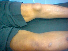
- Swelling over the injury.
- Loss of strength (moderate or severe strain).
- Crepitation ("crackling") feeling and sound when the injured area is pressed with fingers,
- Calcification of the muscle or tendon (visible with X-rays).
- Inflammation of the tendon sheath.
Treatment
Follow your doctor's instructions. Instructions are supplemental.
- Use ice massage 3 or 4 times a day for 15 minutes at a time. Fill a large styrofoam cup with water and freeze.Tear a small amount of foam from the top so ice protrudes. Massage firmly over the injured area in a circle about the size of a softball.
- After the first 24 hours, apply heat instead of ice, if it feels better. Use heat lamps, hot soaks, hot showers, heating pads, or heat liniments and ointments.
- Take whirlpool treatments, if available.
- Wrap the injured knee with an elasticized bandage between treatments.
- Massage gently and often to provide comfort and decrease swelling.
Home Diet
Eat a well-balanced diet that includes extra protein, such as meat, fish, poultry, cheese, milk and eggs. Increase fiber and fluid intake to prevent constipation that may result from decreased activity.
Prevention Tips
- Participate in a stretching, strengthening and conditioning program appropriate for your sport.
- Warm up before practice or competition.
- Tape the knee area before practice or competition.


 Injury to the knee joint in which the upper and lower leg bones are displaced and no longer touch each other. Knee dislocations often include torn or ruptured ligaments in the knee.

 Bruising or tearing of the kidney or ureter. Kidneys filter waste material from the bloodstream and produce urine. Ureters are the tubes that carry urine from the kidneys to the bladder. The most common injury to the kidney is contusion. In contact sports, this may result from a blow from a knee or helmet, with the shock penetrating the flank muscles and reaching the kidney.
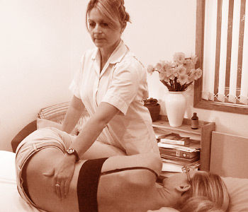 Violent overstretching of one or more ligaments in the temporo-mandibular joint. Sprains involving two or more ligaments cause considerably more disability than single-ligament sprains. When the ligament is overstretched, it becomes tense and gives way at its weakest point, either where it attaches to bone or within the ligament itself. If the ligament pulls loose a fragment of bone, it is called a sprain-fracture. There are 3 types of sprains:
  A complete or incomplete break in the lower jaw (the mandible). The temporo-mandibular joints (TMJ) are located just in front of the ears. These joints connect the lower jaw with the skull and are used to open and close the mouth. A fracture usually occurs at the condyle, or head of the mandible.
 Injury and displacement of the end of the lower jaw from its normal niche in a small depression at the base of the skull.
  Inflammation of the synovium, the smooth, lubricated lining of the hip joint.The synovium's lubricating fluid allows the hip to move freely and prevents bone surfaces from rubbing against each other. Synovitis is often a complication of an injury, such as a fracture, or of collagen diseases, such as gout or rheumatoid arthritis.
  Injury to the muscles and tendons attached to the trochanter, the large end of the femur (thigh bone) that forms part of the hip joint. Muscles, tendons and bone comprise units. These units stabilize the hip joint and allow its motion. A strain occurs at the weakest part of a unit. Strains are of 3 types:
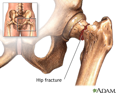 A complete or incomplete break in the head of the femur, the major bone In the hip joint.
 A serious hip injury in which adjoining bones in the hip are displaced so they no longer touch each other. Dislocations are frequently accompanied by bone fractures, torn ligaments and torn tendons. Temporary or permanent damage to bone or to the sciatic nerve makes immediate treatment necessary.
  Inflammation of the bursa surrounding either of the big knobs of bone (trochanters) at the top of the femur (thigh bone). Bursitis may vary in degree from mild irritation to an abscess formation that causes excruciating pain.
  Skull fractures may be of two types:
 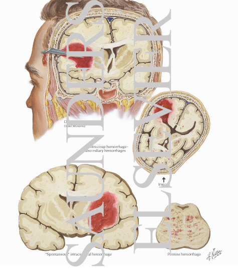 Bleeding (hemorrhage) that causes blood to collect and partially clot (hematoma) inside the brain. The use of CAT scans has shown that this condition occurs more frequently than physicians previously thought.
  Bleeding (hemorrhage) between the skull and the outermost of 3 membranes (meninges) that cover the brain, resulting in a pooling of blood (hematoma) that causes pressure on the brain.
  Bruising of the brain following a blow. Contusions cause bleeding from ruptured small capillaries that allow blood to infiltrate brain tissue.
 A violent jar or shock to the brain that causes an immediate change in brain function, including possible loss of consciousness.
  Inflammation of a tendon (tendinitis) or the lining of a tendon sheath (tenosynovitis) in the hand. This lining secretes a fluid that lubricates the tendon. When the lining becomes inflamed, the tendon cannot glide smoothly in its covering.
  Violent overstretching of one or more ligaments in the hand. Sprains involving two or more ligaments cause considerably more disability than single-ligament sprains. When the ligament is overstretched, it becomes tense and gives way at its weakest point, either where it attaches to bone or within the ligament itself. If the ligament pulls loose a fragment of bone, it is called a sprain-fracture. There are 3 types of sprains: